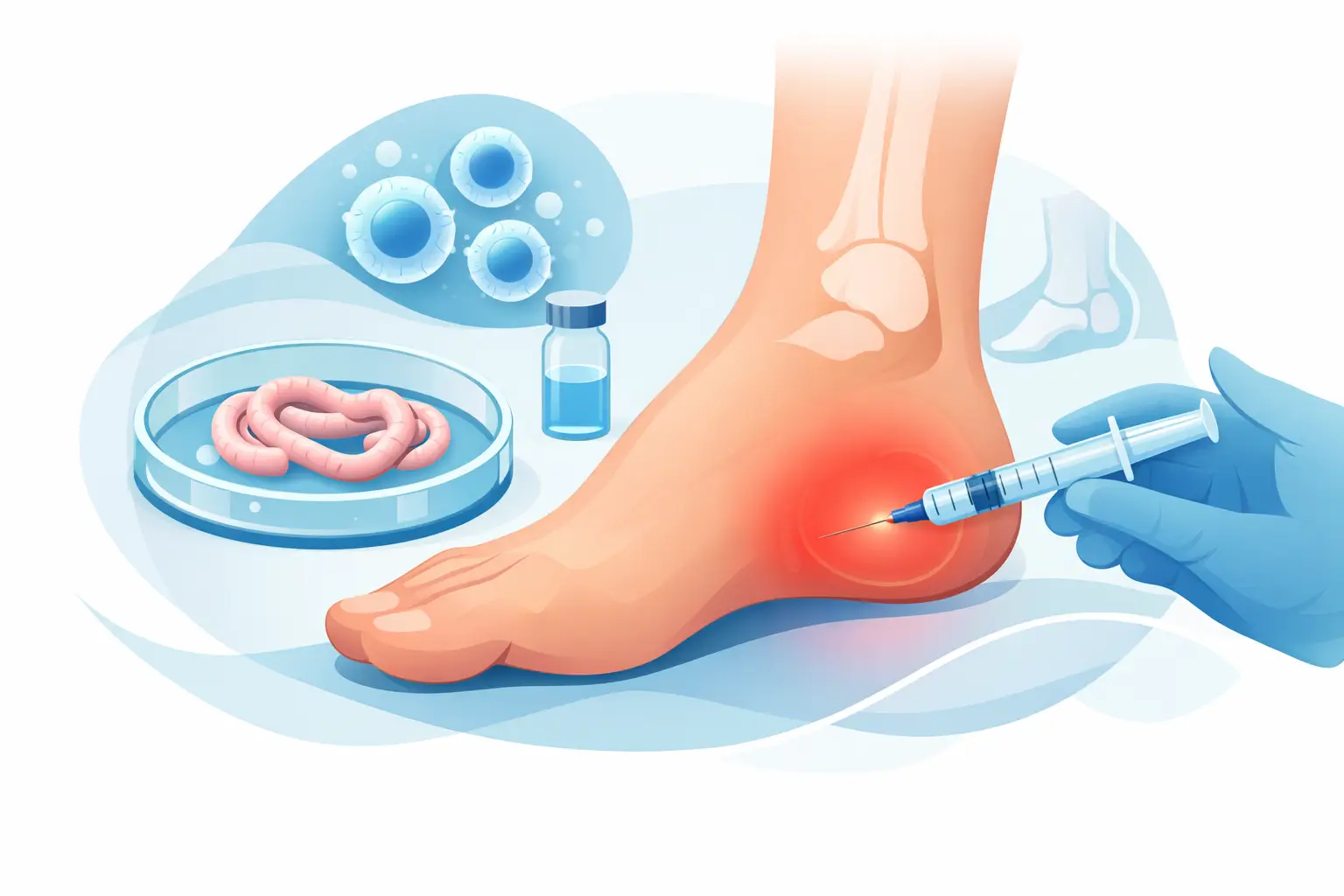

A small cut on the foot can become a much larger problem when pressure, swelling, diabetes, poor circulation, or reduced sensation gets in the way of healing. This foot wound closure case study illustrates why closing a wound is not simply about bringing skin edges together. The goal is to create the safest possible conditions for the tissue to heal while protecting the patient’s mobility and reducing the risk of infection or reopening.
The example below is a representative, de-identified clinical scenario based on common wound-care principles. Every patient’s wound, health history, circulation, and healing capacity are different, so the right treatment plan should always be determined through an in-person evaluation.
Foot Wound Closure Case Study: The Initial Concern
A 67-year-old Mesa-area patient came to the office after noticing a cut on the bottom of the forefoot. He had type 2 diabetes and neuropathy, which meant he had limited feeling in his feet. The injury likely began after stepping on a small object at home, but because it did not hurt, he continued walking on it for several days.
By the time he was evaluated, the wound had become deeper and the surrounding skin was irritated from continued pressure. There was drainage, but no fever or severe systemic symptoms. He had been covering the area with a bandage, hoping it would improve on its own.
This is a common and understandable response, especially when a wound appears small. Yet foot wounds can change quickly. The sole of the foot carries body weight with every step, and pressure can keep an open area from closing even when a patient is doing everything they can at home. For someone with diabetes or neuropathy, a wound may worsen without the pain that would normally signal a problem.
Why Immediate Closure Was Not the First Step
A wound cannot always be safely closed at the first visit. If debris, nonviable tissue, infection, excess drainage, or swelling is present, closing the skin too early can trap bacteria or fluid beneath the surface. That can delay healing and create a more serious complication.
The first priority was a careful examination. The podiatric evaluation included the wound’s location, depth, drainage, surrounding redness, odor, and signs of pressure damage. The patient’s circulation and protective sensation were also assessed. Imaging may be appropriate when there is concern for a retained foreign body, bone involvement, or a deeper injury, while additional testing may be needed based on the wound and the patient’s medical history.
In this case, the wound needed cleaning and selective removal of unhealthy tissue before closure could be considered. The patient was also advised to contact his primary care provider regarding blood sugar management, since elevated glucose can interfere with the body’s ability to heal and fight infection.
The Treatment Plan Focused on Pressure Relief
After cleansing and debridement, the wound was dressed with materials selected for its amount of drainage and condition of the surrounding skin. The care plan also addressed the factor most likely to prevent progress: continued pressure on the bottom of the foot.
The patient was placed in an appropriate offloading device and instructed to limit weight-bearing as directed. Depending on the wound’s location and severity, offloading may involve a surgical shoe, removable walking boot, custom padding, felted foam, crutches, or another method that reduces stress on the injured area. There is no single device that is right for every patient. The best option is one that protects the wound while remaining realistic and safe for the patient to use.
That trade-off matters. A very protective device is not helpful if it increases a patient’s fall risk or is so uncomfortable that it is not worn consistently. A personalized plan considers balance, activity level, home environment, work demands, and the ability to return for follow-up visits.
The patient and family were given clear instructions to keep the dressing clean and dry, avoid walking barefoot, inspect the foot daily, and call promptly for increased redness, warmth, swelling, drainage, odor, fever, or a change in skin color. Because neuropathy can mask pain, visual checks were especially important.
Reassessing the Wound Before Closure
At follow-up, the wound showed less drainage and healthier tissue at the base. The surrounding irritation had improved because pressure was being reduced. There were no concerning signs that would make closure unsafe at that point.
This reassessment stage is essential. Wounds do not follow a calendar. Some can be closed after they are adequately cleaned and stabilized. Others heal better with advanced dressings and secondary healing, meaning the body gradually fills in the wound from the inside out. Larger wounds, wounds with poor blood flow, or wounds with infection may require a different approach altogether.
For this patient, the skin edges could be approximated without excessive tension. Closing a wound under too much tension can compromise blood flow to the edges and increase the chance that it will reopen. After preparing the area and using appropriate local anesthesia, the clinician closed the wound and applied a protective dressing.
The closure was only one part of treatment, not the finish line. The patient continued offloading because putting full pressure on a newly closed plantar wound could cause the repair to fail. He was also scheduled for close monitoring rather than being told to wait until something felt wrong.
What Supported Healing in This Case
Over the next several visits, the incision remained intact and the tissue continued to improve. The patient followed his offloading instructions, attended dressing checks, and involved a family member in daily foot inspections. Sutures were removed only after the area appeared stable and ready.
Several details worked together to support this progress:
- The wound was assessed early enough to identify pressure and tissue damage before a deeper complication developed.
- Unhealthy tissue and contamination were addressed before attempting closure.
- Offloading reduced repeated trauma from walking on the wound.
- Follow-up visits allowed the care plan to change as the wound changed.
- Diabetes and neuropathy were treated as part of the healing picture, not as background details.
Not every patient will need sutures or another form of primary closure. Some foot wounds are better managed open, with specialized dressings, negative-pressure therapy, infection treatment, vascular evaluation, or other advanced care. When healing is delayed, a podiatrist may also consider whether regenerative options are appropriate for the individual situation. The right choice depends on the wound itself and on the patient’s overall health.
Lessons for Patients and Families
The practical lesson from this foot wound closure case study is simple: do not judge a foot wound by pain level or size alone. A painless wound can still be serious, particularly for people with diabetes, neuropathy, circulation concerns, immune-system conditions, or a history of ulcers.
Seek prompt medical attention for a cut, puncture, blister, crack, ulcer, or surgical incision that is not improving. Urgent evaluation is especially important when there is spreading redness, drainage, foul odor, black or blue discoloration, fever, increasing swelling, or a wound that exposes deeper tissue. If you have diabetes, make foot checks part of your daily routine and avoid trying to cut away calluses or dead skin at home.
At Chambers Foot & Ankle, wound care begins with a careful look at the whole person, including health conditions, mobility needs, circulation, activity level, and the practical realities of daily life. Conservative care is often the starting point, but it must be active and closely monitored when a wound is involved.
A foot wound deserves attention before it becomes an emergency. Early evaluation can help protect the tissue, preserve your ability to stay active, and give healing the support it needs.